

Microscopy in Mongolia
veterinary telemedicine in remote regions

A Metamorphosis in Thinking
In 2015 I finally found out what I was missing. I had been working as a professional engineer developing prototype biomedical devices for 4 years and several of my devices had made it to the clinic or operating theater. Engaging and impactful work though it was, the metrics for my success were numbers and the briefings I received were technical specifications. I had gotten into biomedical engineering because of the people it positively impacted, but in my work I found myself farther away from understanding those same people than I was comfortable with.
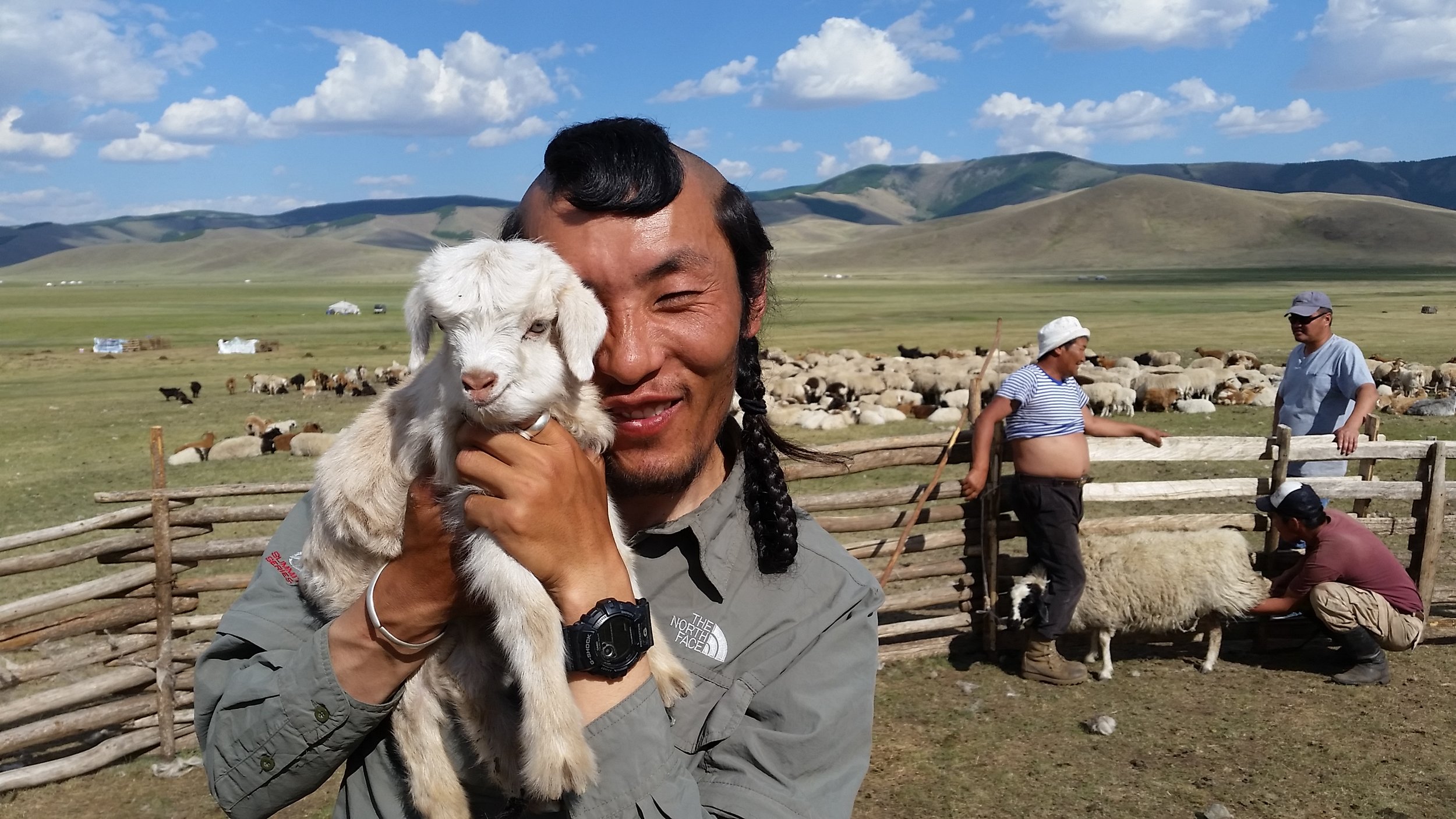
So I decided to do something about it. I sought out projects in my network that would put in in closer contact with users. I was inspired by the emerging need for pathological monitoring in livestock-dependent populations and over nights and weekends I interviewed experts and poured over secondary research. I established myself as expert enough on the topic to write and win a grant from National Geographic to pursue my work in the field in collaboration with the Mongolian Bankhar Dog Project.
Later I would identify many of the tools I used in this project as design thinking and using them was so fundamentally rewarding for their closeness to the people I was making an impact on that I enrolled in the Engineering Design Innovation program at Northwestern University to make human-centered design the center of my career.
Summary
This project aimed to establish the feasibility of the CellScope (on generous loan from the Fletcher lab at UC Berkeley) as a tool for veterinary telemedicine. Specifically, the project aimed to:
Validate the CellScope as an instrument able to image blood films of veterinary species (horses, cattle, sheep, goats and camelids) with diagnostic fidelity comparable to conventional techniques
To evaluate the ability of the CellScope to transmit accurate diagnostic information of blood films taken from livestock to experts from remote field sites in Mongolia.
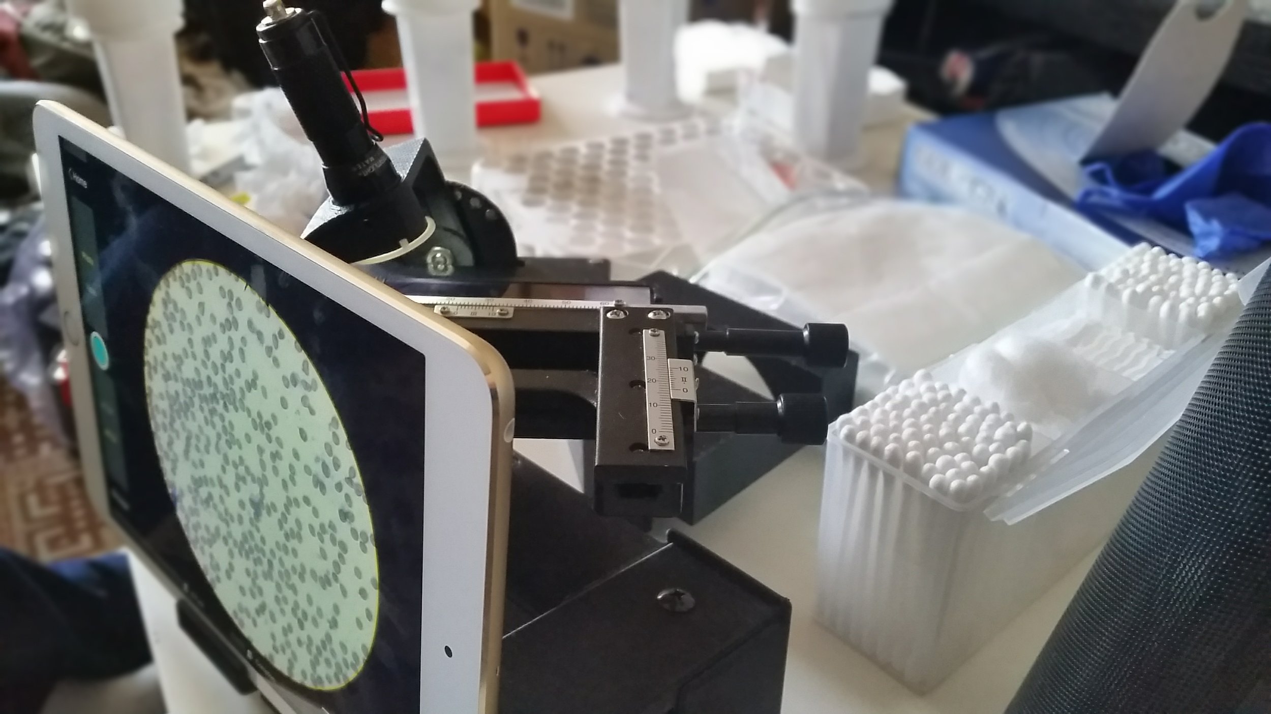
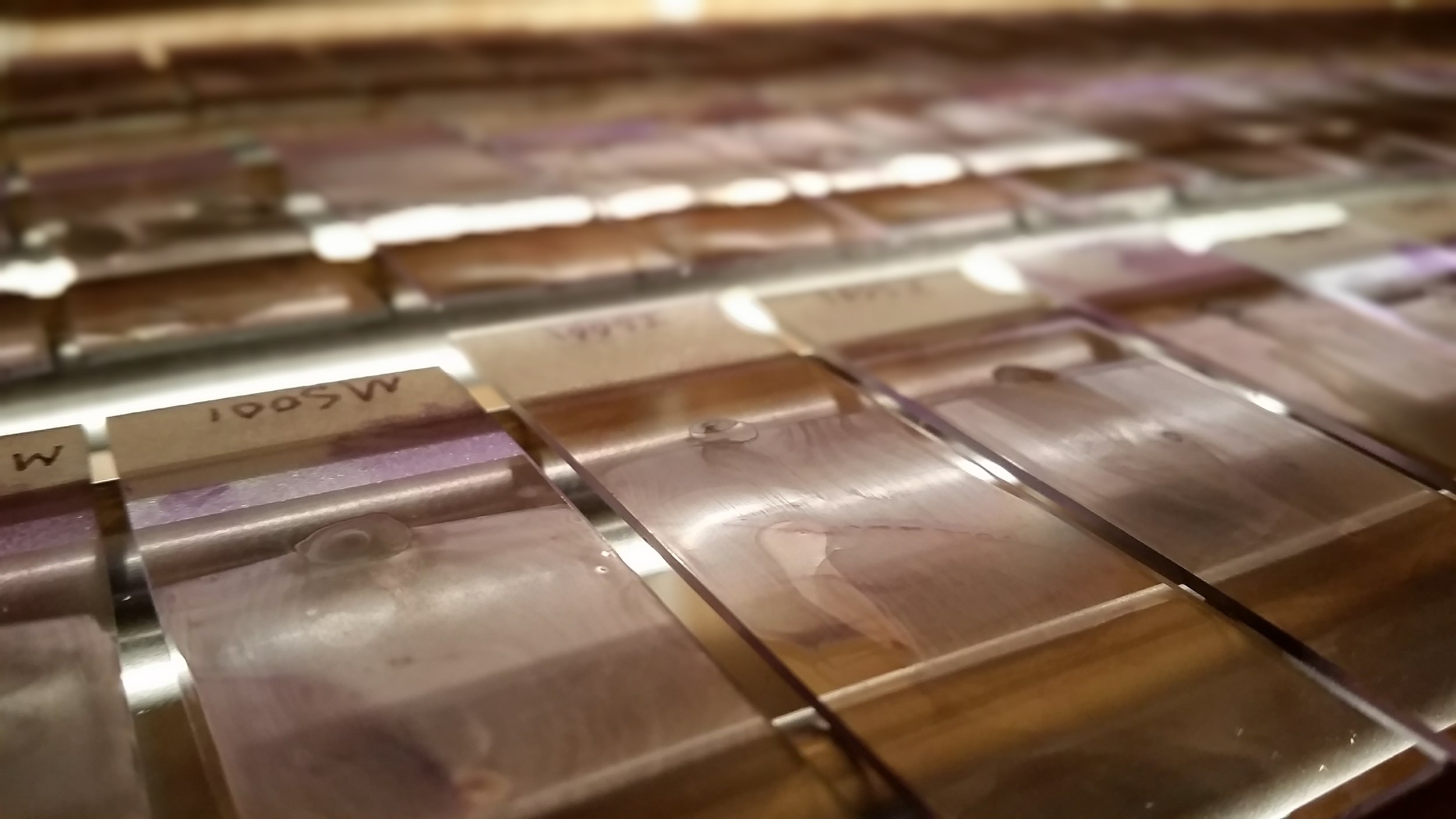
Samples from species relevant to veterinary medicine in Mongolia were collected across multiple sites and ecologies. Slides were prepared from these samples per standard techniques. The slides were imaged by the CellScope and complete blood cell counts (CBC) were performed on the slides by conventional techniques and the image sets of the same slides.
The CellScope was found to be an excellent collector of images and captures almost all the details necessary to perform a CBC. Particularly, leukocyte differentials were difficult, but still possible. Yet, the number of images we acquired would already be prohibitive to the use of the CellScope for remote CBCs since the size of the data to be transmitted would not be supported by current cellular infrastructure in the countries that the CellScope might be used in. That being said, there are a number of other diagnostic tests that can be performed on a manageably small set of images or even a single image. Fecal smears, floats, and sediments, for example, would require a small number of images each. Indeed, we successfully demonstrated the identification of multiple species of parasites using such remotely transmitted images. Furthermore, continued development of the supporting software could potentially make the transmitted data size more manageable for diagnostic tests such as CBCs that might require a larger data set.
In the future, the CellScope could readily see wider deployment to sites in multiple ecologies for early detection of a large variety of diseases relevant to veterinary medicine.
Hypothesis
The CellScope can image blood films of domestic animal species with the fidelity to perform accurate complete blood counts (CBC). These diagnostic images can be acquired and transmitted to hematology and clinical pathology experts to facilitate disease monitoring and/or emerging pathogen surveillance in livestock and wildlife.
Methods
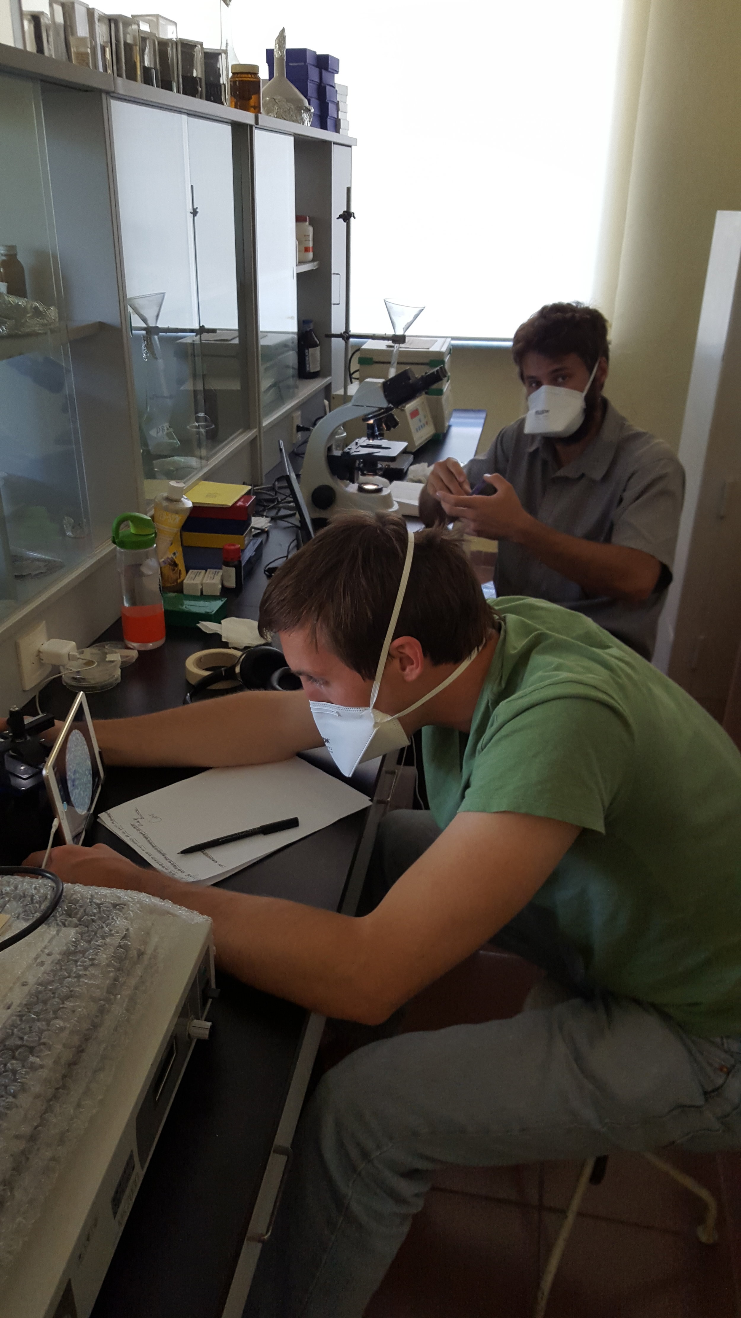
The CellScope was investigated for its feasibility previous to deploying it for field study. Due to the transition from using the originally proposed technology to the CellScope, the parameters of this preliminary investigation had to change. The CellScope imaging platform is immediately familiar to the user of a benchtop microscope. The inventors have already conducted studies characterizing its performance and as such, the metrics that the preliminary investigation would have yielded have already been quantified. Nonetheless, a veterinary pathologist qualified the images taken by the CellScope as of comparable quality to a benchtop microscope and specifically that the images capture all the features relevant to perform a CBC (e.g. nucleus morphology for leukocyte differential).
In the field, 181 samples from horses, cattle, sheep, goats and camelids were collected across sites in Govi-Altai aimag (province), Terelj National Park area, and Hustai National park area. Slides were prepared by smearing a droplet across the slide with a second slide. Slides were allowed to air dry, then were fixed and stained with a modified Giemsa (Diff-quik) kit. Of the total pool, a representative set was selected for imaging and CBC analysis. For each selected slide, images were taken in areas where ~50% of the RBCs were touching, i.e. the readable monolayer, as per standard CBC practice. 10 images were taken of fields that included red blood cells (RBCs) and at least 100 white blood cells (WBCs) were captured in as many images as necessary. 2 images were taken of the feathered edge of the film to confirm slide integrity. The image sets from each of the slides were transmitted to a veterinary pathologist along with the original slides. The pathologist performed CBCs on each the original slides, then performed CBCs based on each of the image sets. Image set analysis order was purposefully uncorrelated to the slide analysis order to prevent bias.
Discussion
The approach and methods were successful in showing that the CellScope can be used for telemedicine applications and exploring the largely unexplored field of remote medicine.
Diagnostic telemedicine, in the broad strokes, focuses on removing a prohibitive step from the process of moving from a sample being collected to a diagnosis being delivered. In Mongolia and other countries with a large livestock-dependent population, veterinary infrastructure can be lacking. The existing personnel are spread too thin and the cost in both time and money make sending any but a minimum of samples out for diagnosis in local or foreign labs make access to veterinary care largely unavailable or impractical. In this project, the CellScope was used to transmit data about the samples for remote diagnoses, thus removing the prohibitive step of needing veterinary personnel to travel long distances to take samples and send them away for diagnosis. In this, the project was successful in demonstrating the ability to transform samples into data that can be used to remotely form a diagnosis. It will be up to larger scale deployment of the CellScope, or likely a future iteration of the device, to determine the broad scale impact of wider usage on the veterinary ecology of the country.
Telemedicine is a growing field and the methods and approaches for attempting to make an actionable diagnosis in the field are still early in their evolution. The usage of the CellScope for telemedicine is also relatively new and how it should be used to capture data relevant to make diagnoses still needs to be codified. For example, the method used in this project for imaging leukocytes can be modified such that the operator uses a randomized scanning pattern to prevent scan bias during image acquisition. Additionally, modifications to the hardware of the CellScope will make it easier to standardize its usage.
Possibly the most important contribution this study presents is to highlight the need for methodological innovation in assessment of telemedicine devices. The methods used in this project were based on the best information available, but methodologies for remote manual CBCs based on image sets have not been much explored. Further study is needed to test its validity or find a better method.